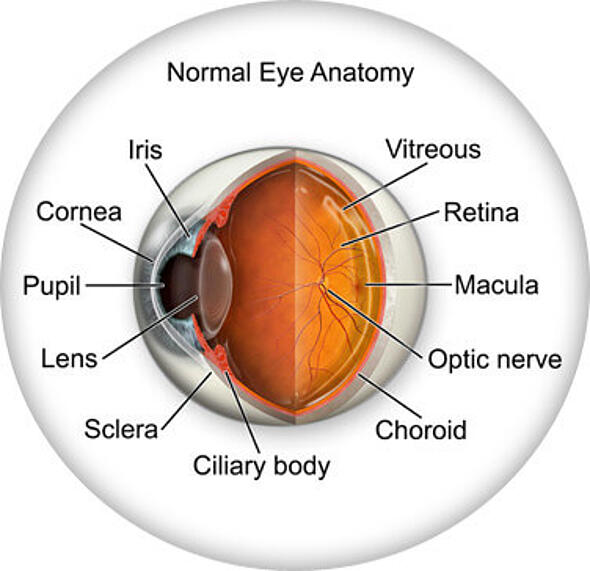
Information About the Eye

Eye Anatomy


The retina:

The retina is the light-sensitive layer of tissue at back of the eye. Similar to the film in a camera, images that come through the lens are focused on the retina. The retina then converts these images into electrical impulses and sends them to the brain. The retina is comprised of photoreceptor cells called rods and cones. There are about 120 million rods in the retina, but only 6 - 7 million cones. Rod receptors are mostly located in the periphery of the eye, are highly sensitive to light, and therefore useful to detect the presence of light. The cone receptors are primarily located in the center of the retina, called the fovea, and produce colored vision and allow seeing details such as reading.

Vitreous body:

The Vitreous body is the clear jelly-like substance that fills the space between the lens and the retina of the eyeball.

Ciliary body:

The ciliary body is made up of the ciliary muscle and the suspensory ligament. This structure functions mainly to hold the lens in place, change the shape of the lens so light can be focused on the retina, and to produce aqueous humor, the fluid that causes the eye to maintain its shape.

Conjunctiva:

The thin and clear membrane that covers the white part of the eye (sclera) and the inner part of the eye lids.

Iris:

The iris is the structure in front of the lens that increases and decreases the size of the pupil to regulate the amount of light entering the eye.

Pupil:

The pupil is the round opening in the center of the iris that lets light in. Pupil size is controlled by the dilator and sphincter muscles of the iris, and regulates the amount of light that enters the eye.

Cornea:

The cornea is a transparent structure that covers the iris and pupil. It is dome shaped and functions similar to an outer lens, by refracting (bending) light. The cornea provides most of our eye’s focusing power.

Lens:

The lens is a transparent structure located between the iris and the vitreous body. The lens is surrounded by a transparent and elastic capsule. It is bi-convex meaning it is rounded on both sides and changes its shape to change the focal distance of the eye. When light enters the eye, the cornea refracts this light, and the lens adjusts its shape and focuses the light upon the retina so that objects appear clearly. As part of the aging process our lenses naturally become less flexible and transparent. This is why we need reading glasses as we age.

Sclera:

The sclera is the white part of the eye that forms 5/6 of the posterior section of the eye.

Choroid:

The choroid is a layer of blood vessels that provide nourishment to the outer layer of the retina. It is an extension of the iris and the ciliary body, located between the retina and the sclera.

Macula:

The macula makes up only a small part of the retina, yet it is much more sensitive to detail than the rest of the retina. The macula allows you to thread a needle or read small print.

Optic nerve:

The optic nerve is located in the posterior section of the eye and carries electrical impulses from photoreceptor cells in the retina to the brain.

Refractive Errors

Perfect-vision Eyes: 

In a normal eye, the incoming light rays are refracted by the cornea and directed through the pupil and focused upon the retina. The retina is made up of light sensitive cells termed rods and cones. These cells transform the light into electrical impulses sent by our optic nerve to our brains.

Myopia:

Myopia or nearsightedness occurs when the eyeball is too long relative to the focusing power of the cornea and lens of the eye. This causes light rays to focus at a point in front of the retina, rather than directly on its surface, so that objects located far in the distance appear blurred. Nearsightedness can be corrected by using a concave lens which causes the light that enters the eye to be focused directly onto the retina.

Hypermetropia: 

Hypermetropia or farsightedness is a vision problem that occurs when light rays entering the eye focus behind the retina, rather than directly on it. This is primarily the case when the eyeball is shorter than normal. This condition can be corrected by a convex lens that focus the light rays directly onto the retina.

Astigmatism:

Astigmatism is a refractive error in which light fails to come to a single focus on the retina to produce clear vision. Instead, multiple focus points occur, either in front of the retina or behind it (or both). Astigmatism is generally caused by an irregularly shaped cornea. Instead of having a symmetrically round shape, the eye is shaped more like the back of a spoon, with one meridian being significantly more curved than the meridian perpendicular to it. Astigmatism requires an additional "cylinder" lens power to correct the difference between the powers of the two principal meridians of the eye, in addition to the concave or convex lenses used to correct nearsightedness or farsightedness.